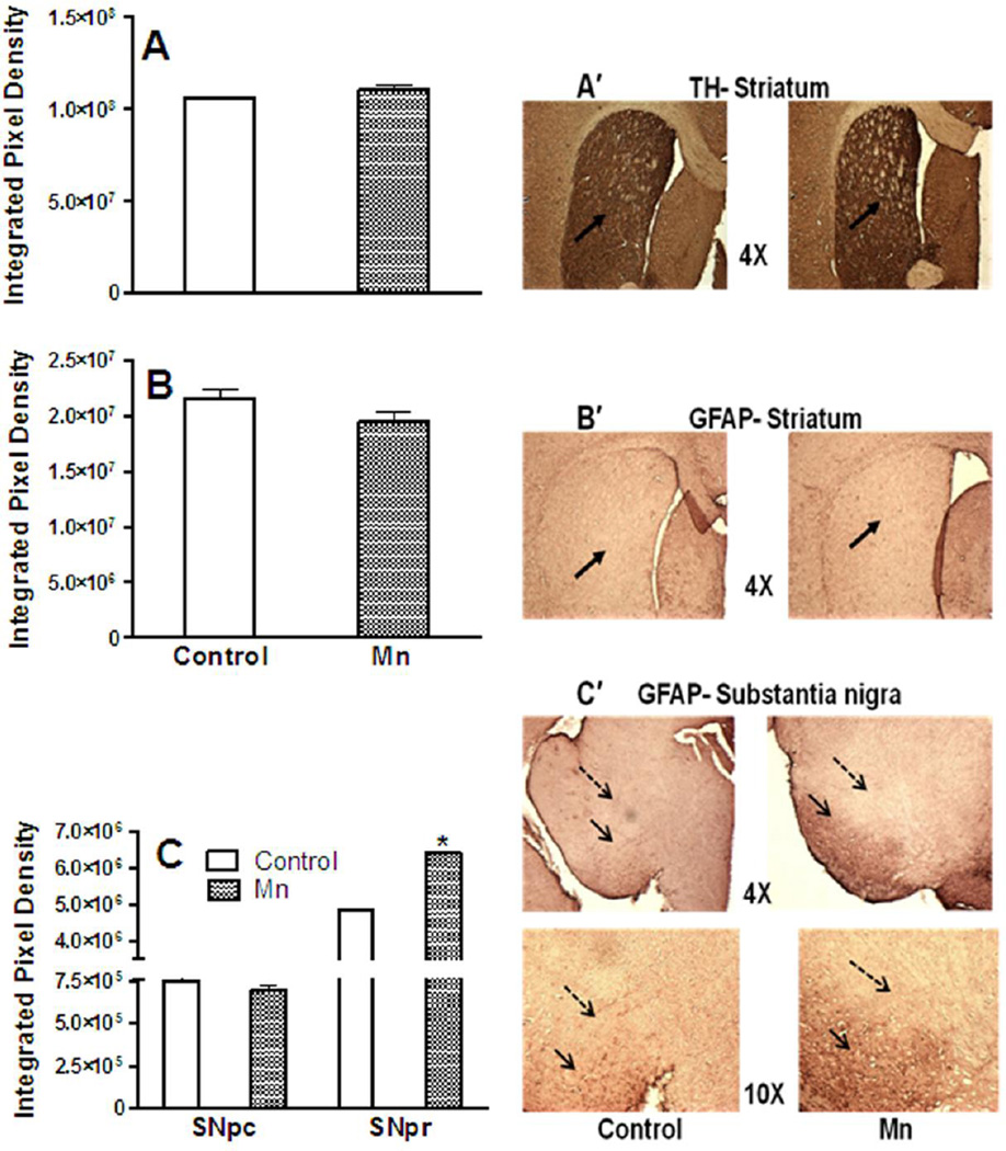
Fig. 9. TH and GFAP Immunoreactivity.
Effect of Mn after 8 weeks of DW (0.4 g/l) exposure on striatal expression of tyrosine hydroxylase (TH; a), striatal expression of glial fibrillary acidic protein (GFAP; b), GFAP expression in the substantia nigra pars compacta (Snpc) and substantia nigra pars reticulata (Snpr; c). Also shown are representative images of striatal TH (a′), striatal GFAP (b′) at 4× magnification and GFAP immunohistochemistry from substantia nigra (c′) at 4× and 10× magnifications. Solid black arrows in images a′ and b′ indicate striatum and images c′ point to Snpr; dashed black arrows in images c′ point to Snpc. Striatal/nigral mean integrated pixel density was used to analyze the intensity of TH and GFAP staining and data are presented as mean ± SEM. * Indicates a significant effect of Mn (p ≤ 0.05).

