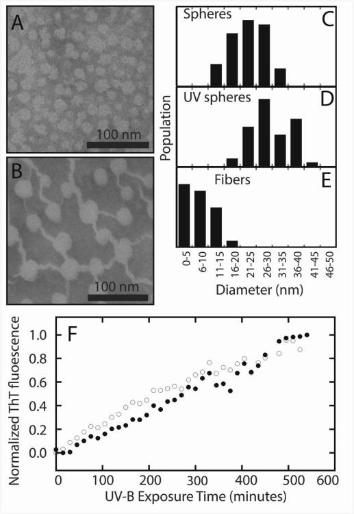
Figure 4.
Aggregates of human γD-crystallin. TEM images of undamaged (A) and UV-B photodamaged (B) γD-crystallin (S84C) show the presence of spherical bodies in both samples, and the formation of fibers in the photodamaged sample after 6 hours of illumination. Analysis of the diameters of spheres and fibers in both samples (C-E) shows a slight increase in sphere sizes from ∼20 nm to ∼30 nm and the formation of fibers with a mean diameter of ∼6 nm. The aggregation of wild type (open circles) and S84C (closed circles) γD-crystallin is accompanied by an increase in ThT fluorescence over the course of 9 hours (F).

