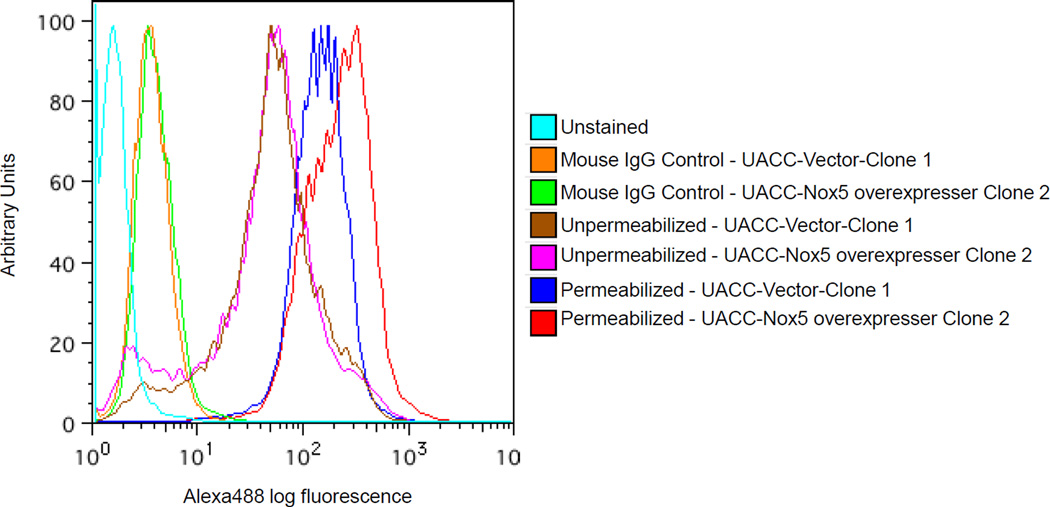
Fig. 6. Flow cytometric detection of Nox5 in UACC-257-Nox5 overexpressing cells.
Log-phase UACC-257 cells that stably express Nox5 (Clone 2) or the vector control (Clone 1) were fixed, permeabilized, and labeled with 5 µg of purified Nox5 mouse monoclonal antibody as described in the Protocol section. Cells labeled with the antibody were staine d with Alexa Fluor 488 goat anti-mouse antibody (1:1000), and the fluorescence was detected by flow cytometry. Unstained cells and cells labeled with irrelevant mouse monoclonal IgG antibody (5 µg) represent background staining controls. Images are representative of at least 3 experiments.

