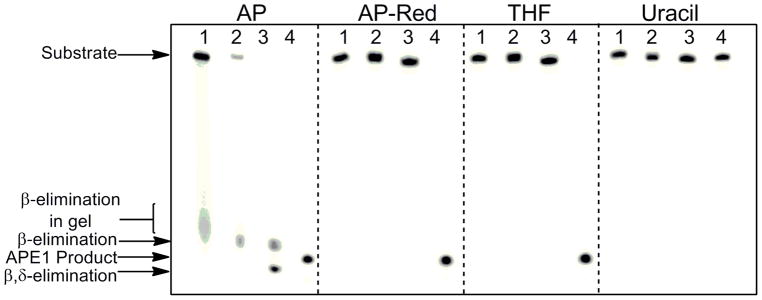
FIGURE 2.

Susceptibility of DNA substrates to β and β,δ-elimination. Autoradiogram revealing β-elimination of authentic AP site before and during gel electrophoresis. Samples contained 30 pmol uracil or THF-containing duplex (annealed to WM complement) in 300 μL of 50 mM HEPES-KOH, 100 mM KCl, pH 7.5 and were incubated with 1.5 units of UDG for 30 min at 37 °C. For AP-Red, uracil-containing DNA and NaBH4 (final concentration of 0.1M) was incubated overnight with 1.5 units of UDG at 37 °C. Following incubation, samples were mixed with an equal volume of Buffer A and subjected to the following: quenching by addition of EDTA and denaturing dye (lanes 1); addition of EDTA followed by in vacuo drying, resuspension in denaturing dye, and heating to 90 °C for 3 min (lanes 2); quenching with 0.1 M NaOH and denaturing dye (lanes 3). All substrates were also allowed to react with APE1, to a final concentration of 500 nM, for 3 min at 37 °C (lane 4). Uracil-containing duplex in the absence of UDG was also used as a control.
