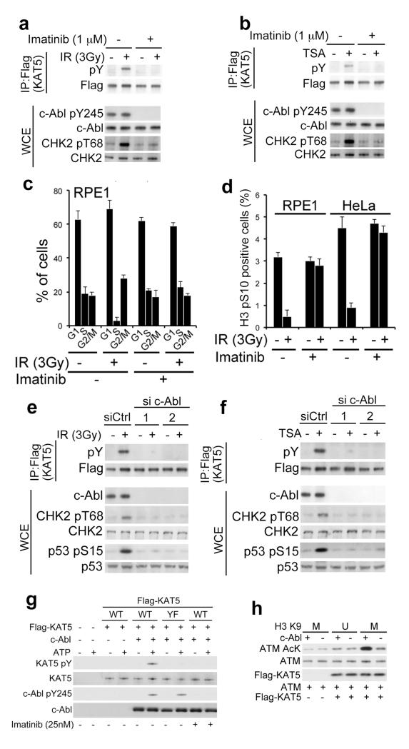
Figure 5. c-Abl-dependent KAT5 tyrosine phosphorylation.
a-b, RPE1 cells were pretreated with imatinib (1μM) for 3 h followed by exposure to IR or TSA; cell extracts were prepared 1 h after IR (a) or 5 h after TSA (b) treatments, and were analysed by immunoprecipitation and western blotting as indicated. c, Flow cytometry analyses of RPE1 cells exposed to IR in the presence or absence of imatinib. Cells were collected 8 h afterwards and analysed. Results are means from three independent experiments ± SE. d, Analysis of G2/M checkpoint in RPE1 and HeLa cells after exposure to IR in the presence or absence of imatinib. Cells were harvested 2 h after IR, stained with H3 pS10 antibody and analysed by flow cytometry. Data from three independent experiments are presented as mean ± SEM. e-f, RPE1 cells were transfected with either of two independent c-Abl targeting siRNAs and, 36 h later were exposed to IR (e) or TSA (f). Extracts were prepared after 1 h and were analysed by immunoprecipitation and western blotting. g, In vitro kinase assays were performed with recombinant c-Abl and purified Flag-KAT5 (WT or YF). Reactions were initiated by ATP addition, and samples were analyzed by western blotting to examine KAT5 tyrosine phosphorylation. h, KAT5 was purified from HeLa cells expressing Flag-KAT5. Purified KAT5 bound to beads was subjected to c-Abl-mediated phosphorylation. After washes, KAT5 was eluted and its activity towards ATM was assessed in the presence of K9 methylated (M) or unmethylated (U) H3 (1-20) peptide by using an AcK antibody.

