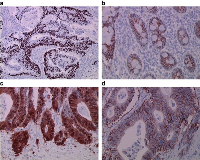
Figure 1.
Representative immunohistochemical staining. (A) Positive p53 immunostaining ( × 200 magnification); (B) in normal small intestinal epithelium, β-catenin was expressed exclusively on the plasma membrane, with no nuclear positivity ( × 400 magnification); (C) tumour cells with aberrant nuclear staining of β-catenin ( × 400 magnification); and (D) tumour cells strongly expressing HER2 (scoring 3+) ( × 400 magnification). Amplification of HER2 was observed in this case.

