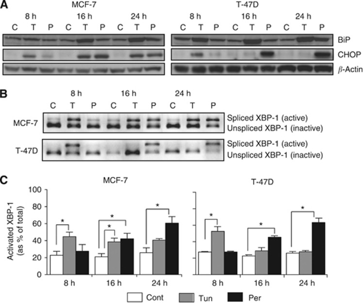
Figure 3.
Persin-induced apoptosis is associated with the upregulation and activation of markers of endoplasmic reticulum stress. (A) MCF-7 and T-47D cells were treated with an apoptotic concentration of persin (P; 10 μg ml−1), the endoplasmic reticulum stress inducer, tunicamycin (T; 10 μg ml−1), or untreated (C) for 8, 16, or 24 h, and levels of BiP and CHOP were determined by immunoblotting. β-Actin was used as a loading control. Representative blots from three independent experiments are shown. (B) MCF-7 and T-47D cells were treated with tunicamycin (T; 10 μg ml−1), an apoptotic concentration of persin (P; 10 μg ml−1) or vehicle control (C) for 8, 16, or 24 h. RNA was then collected, cDNA produced, XBP-1 cDNA amplified using PCR, and the product digested with the restriction enzyme PstI to determine the proportion of undigested (active) to digested (inactive) XBP-1. Representative gels from at least three independent experiments are shown. (C) Densitometric analysis of undigested vs digested XBP-1 PCR product in MCF-7 and T-47D cells treated with tunicamycin (10 μg ml−1) or persin (10 μg ml−1) for 8, 16, or 24 h, and vehicle controls. Bars, mean of three independent experiments±s.e. *P<0.05 for treated cells vs untreated controls.

