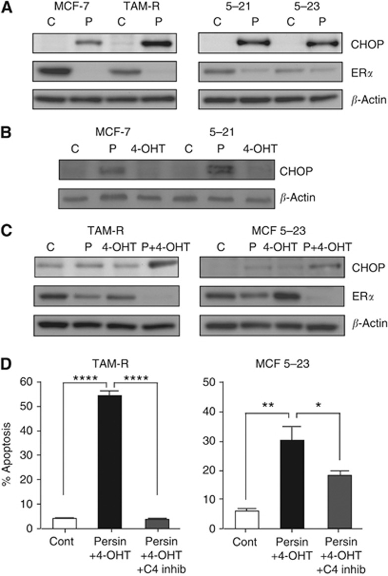
Figure 6.
Persin-induced apoptosis in 4-OHT-resistant cells is associated with changes in markers of endoplasmic reticulum stress. (A) Immunoblot analysis of CHOP and ERα expression in whole-cell lysates from cells treated with an apoptotic concentration of persin (P; 10 μg ml−1) or vehicle control (C) for 24 h. β-Actin was used as a loading control. Representative blots from three independent experiments are shown. (B) Immunoblot analysis of CHOP expression in whole-cell lysates from MCF-7 and MCF 5-21 cells treated with apoptotic concentrations of persin (P; 10 μg ml−1), 4-OHT (4-OHT; 7.5 μM) or vehicle control (C) for 24 h. β-Actin was used as a loading control. Representative blots from three independent experiments are shown. (C) Immunoblot analysis of CHOP and ERα expression in whole-cell lysates from TAM-R and MCF 5-23 cells treated with a non-apoptotic concentration of persin (P; 1 μg ml−1) or 4-OHT (T; 7.5 μM) alone or in combination (P+4-OHT) for 24 h or vehicle control (C) for 24 h. β-Actin was used as a loading control. Representative blots from three independent experiments are shown. (D) TAM-R and MCF 5-23 cells were treated with a non-apoptotic concentration of persin (1 μg ml−1) and 4-OHT (7.5 μM) alone or with caspase-4 inhibitor (20 μM) for 24 h, and then attached and floating populations were analysed for M30-FITC positivity by flow cytometry. Bars, mean of M30-positive fractions from three independent experiments±s.e. *P<0.05; **P<0.005; ****P<0.0001 for combination treatment with inhibitor vs no inhibitor treatment or vehicle controls.

