
Fig. 3.
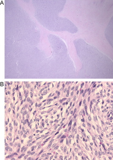
(A) Submucosal low-grade ESS. Pathologic analysis after rectosigmoid resection 2009. HE, 12.5×. Tongues of small blue cells are permeating the muscularis propria. (B). HE, 200×. A diffuse proliferation of spindle cells with little atypia.
Official websites use .gov
A
.gov website belongs to an official
government organization in the United States.
Secure .gov websites use HTTPS
A lock (
) or https:// means you've safely
connected to the .gov website. Share sensitive
information only on official, secure websites.
(A) Submucosal low-grade ESS. Pathologic analysis after rectosigmoid resection 2009. HE, 12.5×. Tongues of small blue cells are permeating the muscularis propria. (B). HE, 200×. A diffuse proliferation of spindle cells with little atypia.