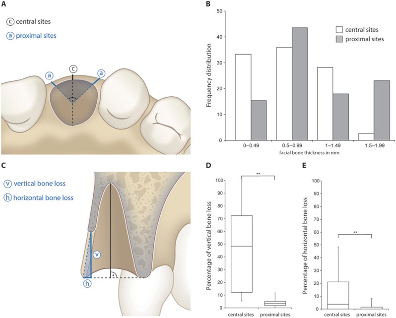
Figure 2.
Baseline measurements and dimensional and vertical bone loss after 8 wks of healing. (A) The analysis was performed in central (c) and proximal sites (a) oriented at a 45° degree angle with the tooth axis as a reference. (B) Frequency distribution of facial bone wall thickness in central and proximal sites. (C) A horizontal reference line was traced connecting the facial and palatal bone wall for standardized measurements. The point-to-point distance between the 2 surface meshes with the respective angle to the reference line was obtained for each sample, and the vertical and horizontal bone losses were calculated accordingly. (D) Percentage of vertical bone loss in central and proximal sites. (E) Percentage of horizontal bone loss in central and proximal sites. **p < .0001.

