
Figure 1.
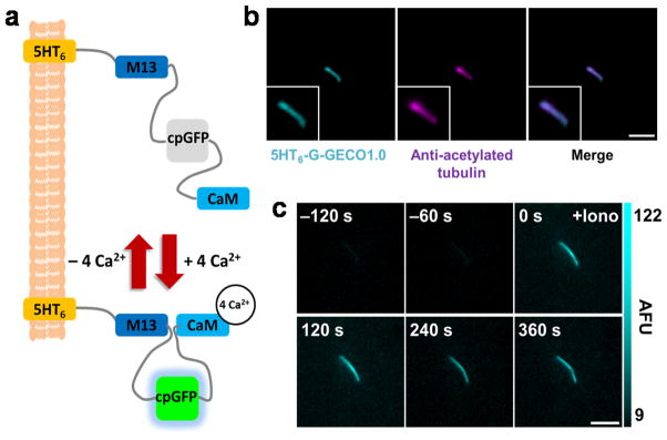
5HT6-G-GECO1.0 targets primary cilia and detects changes in ciliary Ca2+. (a) Schematic of 5HT6-G-GECO1.0. G-GECO1.0 contains M13 (a skeletal muscle light-chain kinase), a circularly permuted GFP (cpGFP), and Calmodulin (CaM). (b) A primary cilium from a NIH-3T3 cell expressing 5HT6-G-GECO1.0 stained with antibody against acetylated α-tubulin. Bar represents 3 μm. (c) Time-lapse imaging of a NIH-3T3 primary cilium expressing 5HT6-G-GECO1.0 treated with ionomycin (Iono). AFU stands for arbitrary fluorescence unit. Bar represents 5 μm.