Fig. 1.
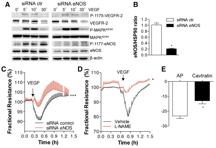
eNOS activity is crucial for VEGF-induced permeability in HDEMCs. (A) HDMECs with control or eNOS siRNA were stimulated with vehicle (control, 0 minutes) or VEGF (100 ng/ml) for 5, 10 and 30 minutes and the cell lysates were analyzed by western blotting for eNOS, P-eNOS1177, P-1175-VEGFR-2, VEGFR-2, P-MAPK42/44, MAPK42/44 (where P indicates the phosphorylated form). (B) Quantification of eNOS levels in HDEMCs by densitometry from four independent experiments and expressed relative to the amount of β-actin. The data shown represent the mean±s.e.m. from five individual experiments. *P<0.05 compared to HDEMCs with siRNA control. (C) TEER measurements of post-confluent HDEMCs transfected with control (black line) or eNOS (red line) siRNA expressed as fractional resistance (%) of the TEER basal values. HDEMCs were stimulated with VEGF (100 ng/ml; addition indicated by the arrow) and TEER measured overtime. *P<0.05 compared to HDEMCs with control siRNA. (D) TEER measurements of HDEMCs treated with L-NAME (1 mM) or vehicle followed by VEGF (100 ng/ml) stimulation. *P<0.05 compared to vehicle-treated HDEMCs. (E) TEER measurements of HDEMCs treated with the eNOS inhibitor cavtratin peptide (10 µM) or the peptide control (antennapedia internalization peptide, AP) for 1 hour, measured after VEGF stimulation for 5 minutes. *P<0.05 compared to AP-treated cells (n = 3). The data shown represent the mean±s.e.m. from three individual experiments.

