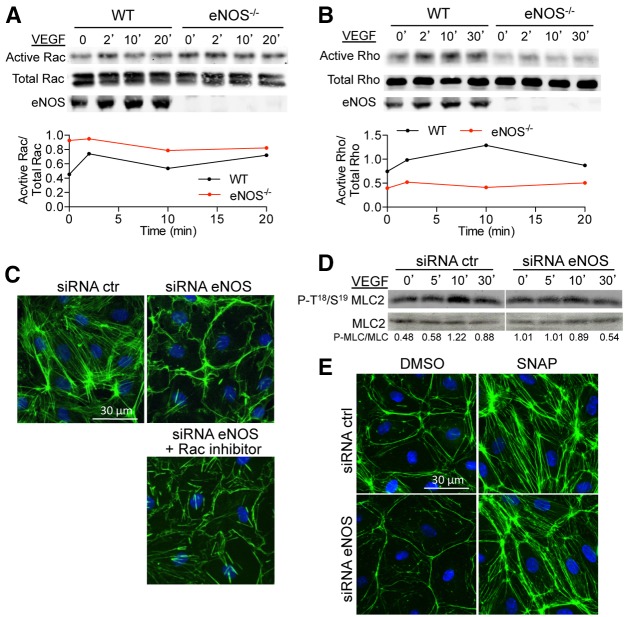
Fig. 3.
eNOS deficiency enhances Rac activity while reducing Rho activity. (A) WT and eNOS−/− MLECs were stimulated with VEGF (100 ng/ml) for the indicated times and Rac activity was measured as described in the Materials and Methods. Total Rac and eNOS levels were determined in the whole cell lysates before GST–PAK incubation. The graph shows the densitometric ratio of active Rac to total Rac. The data are representative of three independent experiments. (B) WT and eNOS−/− MLEC were stimulated with VEGF (100 ng/ml) for the indicated times and cell lysates processed for Rho activity. Total Rho and eNOS levels were determined in the whole cell lysates before GST–Rhotekin incubation. The graph shows the densitometric of active Rho to total Rho. The data are representative of three independent experiments. (C) HDEMCs with control or eNOS siRNA were pre-incubated with Rac inhibitor (25 µM, 6 hours) and stimulated with VEGF (100 ng/ml) following immunolabeling of F-actin (green) and nuclei (blue). (D) Western blotting for P-T18/19 MLC2 and MLC2 in whole cell lysates from HDEMCs with eNOS or control siRNA stimulated with VEGF (100 ng/ml) for the indicated times. The data represent three independent experiments. (E) HDEMCs with eNOS or control siRNA were stimulated with vehicle or SNAP, an NO donor (100 µM, 1 hour) and immunolabeled for F-actin (green) and nuclei (blue). Images are representative of three experiments.

