Fig. 1.

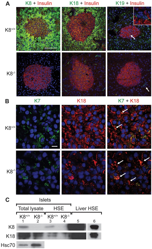
K8 and K18 are main keratins in the endocrine pancreas of K8+/+ mice but low levels of K7 also exist, allowing marginal keratin filament formation in K8−/− mice. (A) Immunostaining of pancreatic sections from K8+/+ and K8−/− mice for insulin (red), K8, K18 or K19 (green) and nuclei (blue). K8 and K18 are the most prominent keratins in the islets and some K19-positive cells are seen in K8+/+ islets. The white arrow in the upper panel points to K19-positive cells and the inset shows a higher magnification of the K19-positive and insulin-negative cells that form occasional duct-like structures within the islet in K8+/+ mice. K8−/− mice lack K8 but express K18 in islets and K19 in exocrine ducts (indicated by the white arrow in the lower panel). Scale bar: 100 µm. (B) Islet cells in pancreatic sections from K8+/+ and K8−/− mice stained for K7 (green), K18 (red) and nuclei (blue). Arrows show colocalisation of K7 and K18. Scale bar: 10 µm. (C) Western blot for K8, K18 and Hsc70 of islet total lysates (lane 1, K8+/+; lane 2, K8−/−), for K8 and K18 of HSE islet lysate (lane 3, K8+/+; lane 4, K8−/−) and liver HSE (lane 5) used as positive control. Lane 6 shows a shorter exposure of the K8 and K18 signals seen in lane 5.
