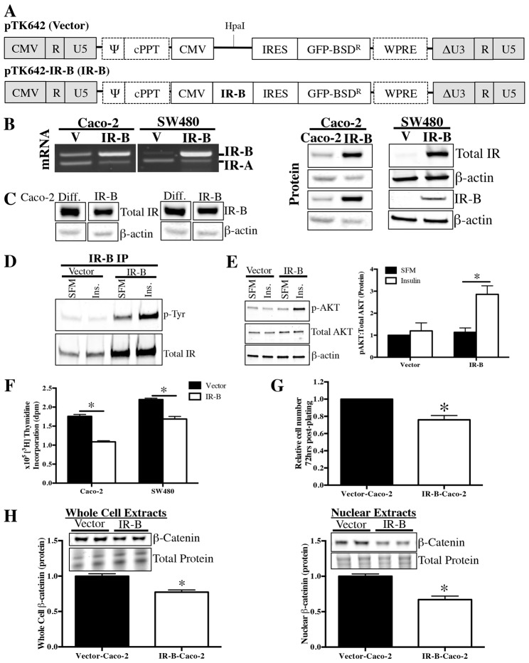
Fig. 6.
Stable overexpression of IR-B in subconfluent Caco-2 cells attenuates cell proliferation. (A) Lentiviral constructs were used to generate Caco-2 and SW480 cells with stable, predominance of IR-B expression (IR-B) or empty vector control (V). The remaining viral components of the vector were the long terminal repeats (LTR), virus packaging signal (Ψ) and the polypurine tract (cPPT). Expression of viral vector components was mediated by the 5′ CMV promoter and polyadenylation signal. The expression cassette contained a CMV promoter, human insulin receptor isoform B (IR-B) gene (in IR-B only), and internal ribosomal entry site (IRES) mediating expression of green fluorescent protein fused to a blasticidin-resistance gene (GFP-BSDR) and Woodchuck hepatitis virus post-transcriptional regulatory element (WPRE). The U3-deleted self-inactivating sequence (ΔU3) was in the 3′LTR. Vector backbone was previously described (Titus et al., 2012). (B) RT-PCR with primers spanning exon 11 confirms stable, predominant IR-B expression at the mRNA level. Western blot for total IR and IR-B in subconfluent vector and IR-B cells confirm enhanced IR-B protein levels. Similar β-actin levels confirm equal loading. (C) Western blot confirms lentivirus-induced IR-B expression levels in subconfluent IR-B-Caco-2 cells approximate, but do not exceed, endogenous increases in total IR or IR-B that occur following differentiation. Samples were all run on the same gel and normalized to β-actin. (D) Immunoprecipitation and western blot to assess tyrosine phosphorylation of IR-B in serum-deprived, subconfluent Vector-Caco-2 and IR-B-Caco-2 cells following treatment for 5 minutes with 200 ng/ml insulin or serum-free medium (SFM) alone. Data represent images of two independent blots. (E) Western blot to assess phosphorylation of AKT following a 5-minute treatment with 200 ng/ml insulin or SFM alone in Vector-Caco-2 and IR-B-Caco-2 cells. β-actin confirmed equal loading and image shown corresponds to p-AKT blot. Loading was identical for total AKT. Samples were run in triplicate. (F) [3H]thymidine incorporation measured DNA synthesis at 72 hours post plating in Vector or IR-B (Caco-2 or SW480) cells. Date represent mean ± s.e.m. of two independent experiments with six replicates each. *P≤0.05 versus Vector, paired t-test. (G) Cell number was assessed at 72 hours post-plating in Vector versus IR-B-Caco-2 cells. Data represent mean ± s.e.m. for two independent experiments performed in triplicate. *P≤0.05 versus Vector-Caco-2, unpaired t-test. (H) Western blotting with whole cell and nuclear extracts measured total and nuclear β-catenin levels in subconfluent Vector and IR-B-Caco-2 cells. Data represent at least two independent experiments performed in duplicate; *P≤0.05 versus Vector-Caco-2, unpaired t-test.

