
Fig. 4.
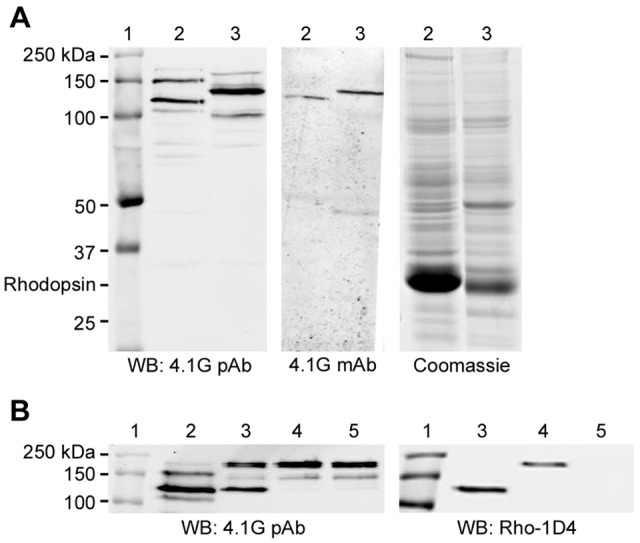
Specificity of 4.1G polyclonal and monoclonal antibodies. (A) Western blots of bovine ROS extracts and mouse retinal membranes. The left panel was labeled with the 4.1G polyclonal antibody (pAb) which detected two dominant variants (125 and 150 kDa) in the bovine ROS (lanes 2) and one dominant (140 kDa) in the mouse retina (lanes 3). The middle panel shows a western blot labeled with the 4.1G monoclonal antibody (mAb) which detected only the dominant variant in the bovine (125 kDa) and mouse (140 kDa) retina. The Coomassie-Blue-stained gel (right panel) is shown for the bovine and mouse samples. Molecular markers were loaded as reference (lane 1). (B) Western blots labeled with the 4.1G polyclonal antibody comparing the endogenous 4.1G variants in the bovine ROS with the cloned 4.1G exogenously expressed in HEK-293 cells. Lane 1, molecular markers; lane 2, lysate of bovine ROS; lane 3, cell lysate transfected with short bovine 4.1G; lane 4, cell lysate transfected with middle bovine 4.1G; lane 5, mock transfection. Note that the short bovine 4.1G (734 aa, lane 3) was of similar molecular mass to the most dominant variant in the bovine retina (lane 2), which were both ∼125 kDa. The 4.1G antibody also detected the endogenous 4.1G in all HEK-293 cell samples, including the mock-transfected cell lysate (∼200 and 150 kDa, lane 5). An immunoblot labeled with Rho-1D4 antibody showed the expression of the 4.1G variants.