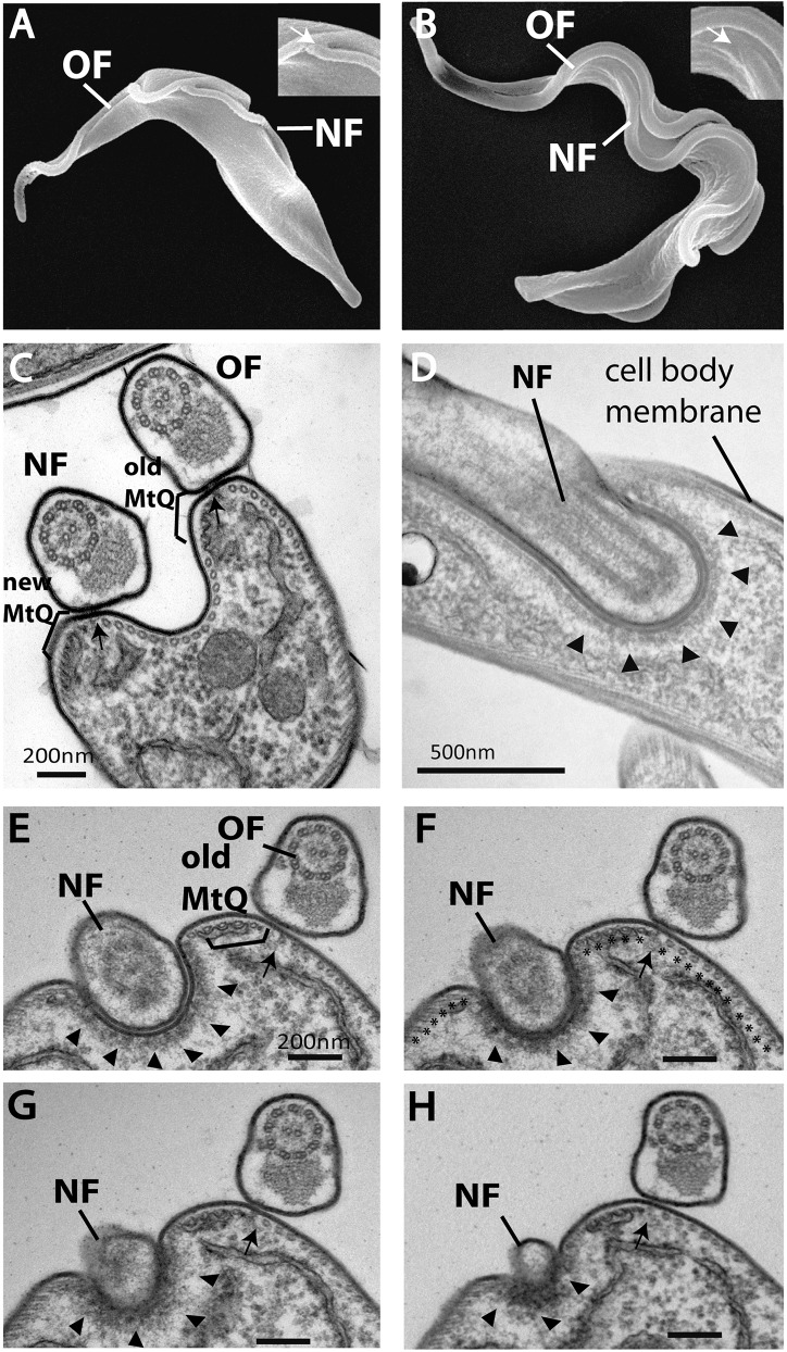
Fig. 1.
The distal tip of the new flagellum in bloodstream form cells is located in an indentation of the cell body membrane. (A) A SEM image of the procyclic form with an old (OF) and new flagellum (NF). The distal tip of the new flagellum is attached to the old flagellum (arrow in inset). (B) A SEM image of the bloodstream form illustrating the distal tip of the new flagellum lying alongside the old flagellum (arrow in inset). (C) A TEM image showing a cross section through an old and new flagellum illustrating the organization of flagella attachment to the cell body through a FAZ filament (arrow) and MtQ (brackets). (D) Longitudinal TEM thin-section through the distal tip of a new flagellum, illustrating the tip embedded within the cell body (arrowheads). (E–H) Adjacent serial cross sections (∼100 nm thick) through an old flagellum and the distal tip of a new flagellum, illustrating the distal tip located in an indentation within the cell body (arrowheads). The old MtQ (bracket) and old FAZ filament (arrow) are indicated in E. Microtubules are shown by asterisks in F.

