
Fig. 6.
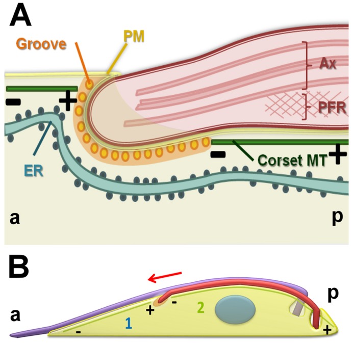
A diagrammatic representation summarizing our interpretation of groove ultrastructure and subpellicular microtubule remodeling. (A) Longitudinal view showing the distal tip of the new flagellum embedded in a groove. Orientation is as indicated (a, anterior and p, posterior). Microtubules terminate with their plus ends anterior to the groove (+). The plasma membrane (PM), axoneme (Ax) and paraflagellar rod (PFR) are shown. (B) Diagram representing a hypothetical remodeling of microtubules around the groove through disassembly (deploymerization or severing) of the plus ends of microtubules located anterior to the tip of the new flagellum (1). The new flagellum is elongating in the direction of the red arrow. Assembly and growth of microtubules posterior to the groove might occur at the minus end immediately posterior to the groove or at the plus end, which is at the posterior end of the cell (2).