
Figure 5.
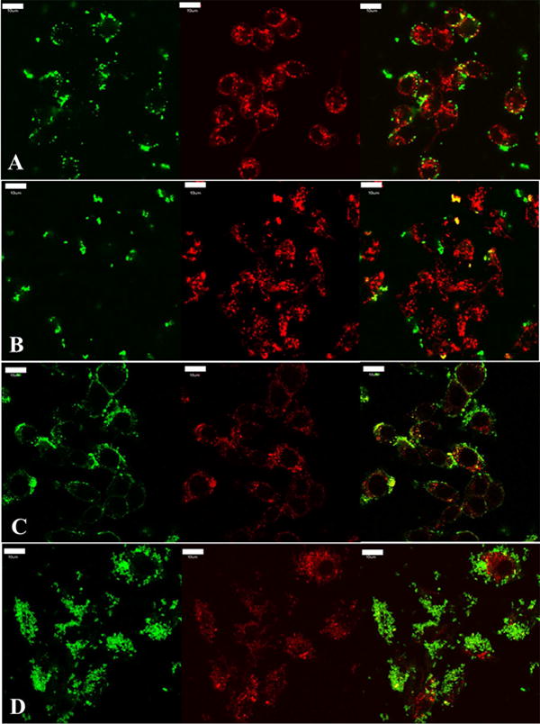
Transferrin and dextran, intracellular markers of clathrin mediated endocytosis and fluid phase endocytosis, respectively, were co-incubated with silica nanoparticles to investigate the degree of co-localization to confirm clathrin and fluid phase mediated mechanisms; RAW 264.7 cells shown. A and B) Depicted is a single focal plane and the respective channels of live cells after 15 mins of incubation. Transferrin is labeled in red, nanoparticles are labeled in green (A is spherical treatment and B is worm like treatment both at 75 μg/mL), and the co-localization is depicted in yellow between particles and transferrin. C and D) Depicted is the fluorescence from all Z stacks and respective channels of fixed cells after 30 mins of incubation. Dextran is labeled in red, particles are labeled in green (C is spherical treatment and D is worm like treatment at 75 μg/mL), and yellow represents the co-localization between nanoparticles and dextran. There appears to be a higher degree of co-localization with both these cellular internalization markers with spherical nanoparticles when compared to worm like nanoparticles, suggesting that a higher degree of clathrin and fluid phase mediated endocytosis occurs with these systems. It is important to note that the co-localization (yellow) in all images, suggests that clathrin and fluid phase mediated endocytosis is at play for all geometries tested. For clarity and due to the significant similarities of worms and cylinders, cylindrical data has been moved to the supplemental materials. Scale bar 10 μm.