
Figure 6.
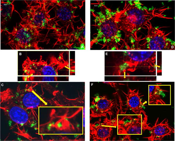
Actin polymerization staining in RAW 264.7 cells, involved in mechanisms of endocytosis is depicted to visualize the hallmark endocytic process involved in the internalization of these particles. Red: phalloidin stain of actin polymerization, green: nanoparticle FITC attachment, and blue: DAPI nucleus stain. A, B and C) Spherical nanoparticle and D, E and F) worm nanoparticle treatment after 15 mins. Spherical nanoparticle treatment (A) and worm nanoparticle treatment (D) appears to induce very different polymerization patterns. The polymerization patterns observed in treated cells include invaginations within the membrane associated with nanomaterials, identified in the zoomed insert in C and in the depiction of the z stack in B. While other polymerization patterns appear to be extravasations from the membrane associated with nanomaterials, marked in the zoomed inserts in F and the z stack in E. These polymerization patterns could suggest the involvement of clathrin mediated, macropinocytic and phagocytic mechanisms.). For clarity and due to the significant similarities of worms and cylinders, cylindrical data has been moved to the supplemental materials.