
Figure 7.
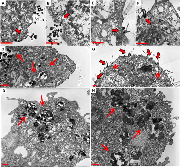
Following 15 mins of incubation with both worm and spherical nanoparticles, cells were fixed and imaged via TEM. Membrane invaginations are associated with spherical particles (A and B). Membrane extravasations however are observed to be associated with worm particles (E and G) and membrane wrapping associated with worm like particles (F and G) are observed. Both nanoparticles are observed within the cytoplasm of these cells at 15 mins (C and G) while there is a greater degree of uptake of spherical nanoparticles at this time point. This suggests that spherical particles are taken up more rapidly than worm nanoparticles. However, a greater amount of both nanoparticles are internalized at 24 hour time points, as we observed previously (D and H).25 Additionally, there appears to be some type of sequestering mechanism at play.). For clarity and due to the significant similarities of worms and cylinders, cylindrical data has been moved to the supplemental materials. Scale bars 1μm.