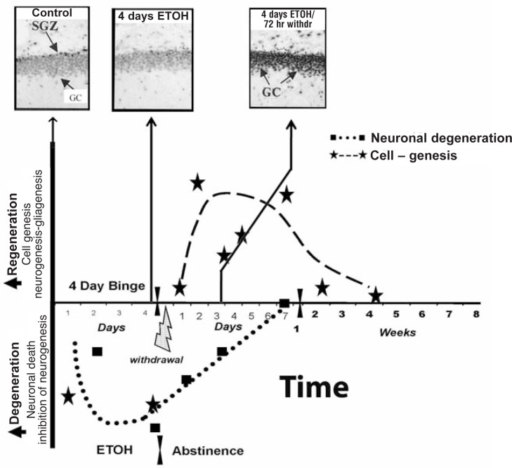
Figure 6.
Regeneration of brain is related to increased phosphorylated cAMP-responsive element-binding protein (pCREB). The 4-day rat BIBD model time line illustrates the relationship between alcohol-induced degeneration, abstinence-induced neurogenesis, and pCREB. The temporal relationship of binge-induced neurodegeneration and abstinence-induced cell genesis can be examined by pCREB immunohistochemical staining in the dentate gyrus. Immunohistochemical staining is a process of localizing proteins in cells of a tissue section using antibodies that bind to specific proteins, such as pCREB. More staining means more protein. In the dentate gyrus granule cells (GCs) of control subjects, most neuronal nuclei have some pCREB+ immunoreactivity (IR), with higher levels of staining in the subgranule zone (SGZ), where neurogenesis is active (control, upper left image). In the diagram, values below the x-axis reflect degeneration or loss of brain mass. Markers of neuronal death increase throughout the 4 days of intoxication. Neurogenesis decreases and pCREB+IR is low (middle image). Markers of neuronal death persist into abstinence, although they progressively decline and mostly disappear after 1 week of abstinence (dotted line). Regeneration is represented by the dashed line increasing above the x-axis, with stars indicating time points of measured neurogenesis and other cell genesis (Crews and Nixon 2008). After 4 days of binge alcohol treatment, pCREB staining is decreased when neurogenesis is inhibited and granule cells degenerate. However, after 72 hours of abstinence, a marked increase in pCREB staining (top photo [4 days alcohol/72 hours withdrawal]) coincides with increased neurogenesis and loss of degeneration markers (Bison and Crews 2003).
NOTE: CREB is a transcription factor altered by alcohol. When CREB is activated, pCREB is formed. The dentate gyrus is part of the hippocampus.

