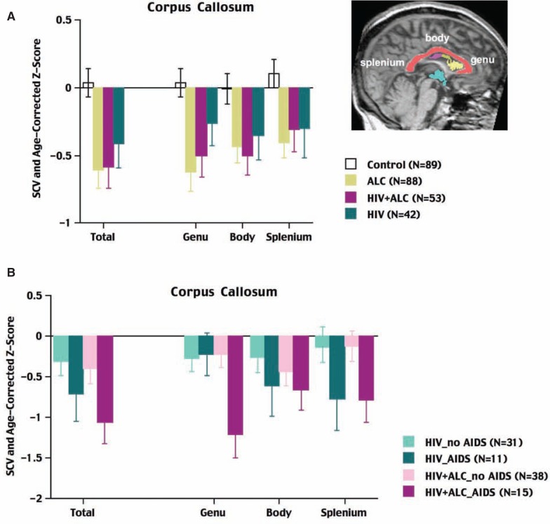
Figure 2.
Magnetic resonance imaging (MRI) of the corpus callosum of the brain. MRI represents a sagittal section along the midline of a brain, with the corpus callosum—the bundle of fibers that connects the right and left halves (i.e., hemispheres) of the brain—displayed in salmon color. Also highlighted in this image are views of the frontal (yellow) and middle (magenta) regions of the ventricles as well as the third ventricle (turquoise) shown in Figure 1A. A) The volumes of the total and regional divisions of the corpus callosum in control subjects, patients with alcoholism only, patients with human immunodeficiency virus (HIV) and alcoholism, and patients with HIV infection only. All three patient groups had smaller volumes of the corpus callosum than the control group, but the abnormalities of the two groups that included alcoholic patients were especially prominent. B) Total and regional volumes of the corpus callosum for HIV-infected patients with and without a history of acquired immune deficiency syndrome (AIDS) and with and without comorbid alcoholism. HIV-infected patients with AIDS and alcoholism showed the greatest callosal abnormalities.
NOTE: The volumes of the three patient groups were adjusted for variation in head size and age of the control subjects; the bars and error bars signify the means ± standard error for each group for each measure.

