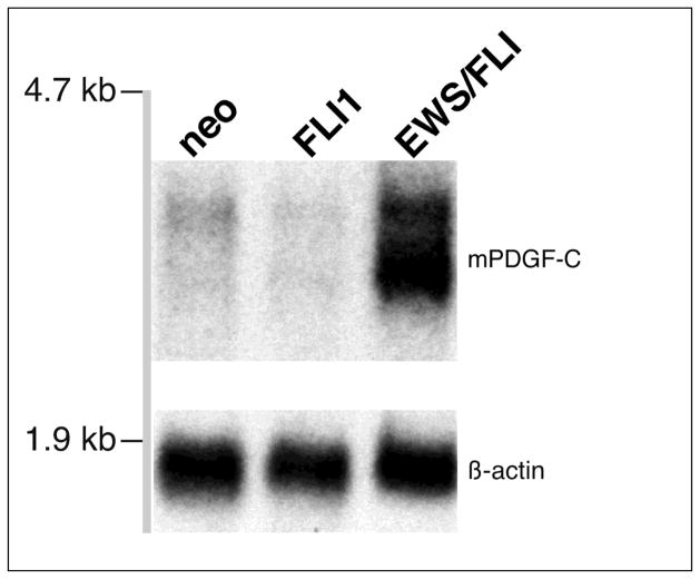
Figure 1. PDGF-C is induced in response to EWS/FLI expression in NIH 3T3 cells.
Abundant PDGF-C expression is detected by northern blot analysis of total RNA from polyclonal NIH 3T3 cells stably expressing the EWS/FLI fusion gene from a retroviral vector. Only low-levels of PDGF-C are apparent in NIH 3T3 cells stably expressing either empty retroviral vector (neo) or the native FLI-1 gene. Probes used are as indicated.

