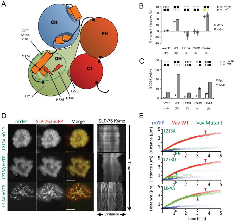
Figure 5. The selective inactivation of the Vav1 GEF active site does not alter microcluster persistence or movement.
(A) The domain structure of Vav mutants used in this figure. (B–C) J.Vav cells were transiently transfected with the indicated constructs. CD69 upregulation (B) and calcium responses (C) were measured as in Figure 2. (D) JV.SC cells transiently transfected with the indicated Vav1.mYFP constructs were stimulated and imaged as in Figure 2B. Representative maximum-over-time projections (left) and kymographs (right) were selected from 3 independent experiments. Scale bars as in Figure 2B. (E) Composite kymographs depicting SLP-76 MC movement and persistence were prepared as in Figure 2C. Arrowheads correspond to the half-life of SLP-76 MC for each condition. See Table 1 for further analysis.

