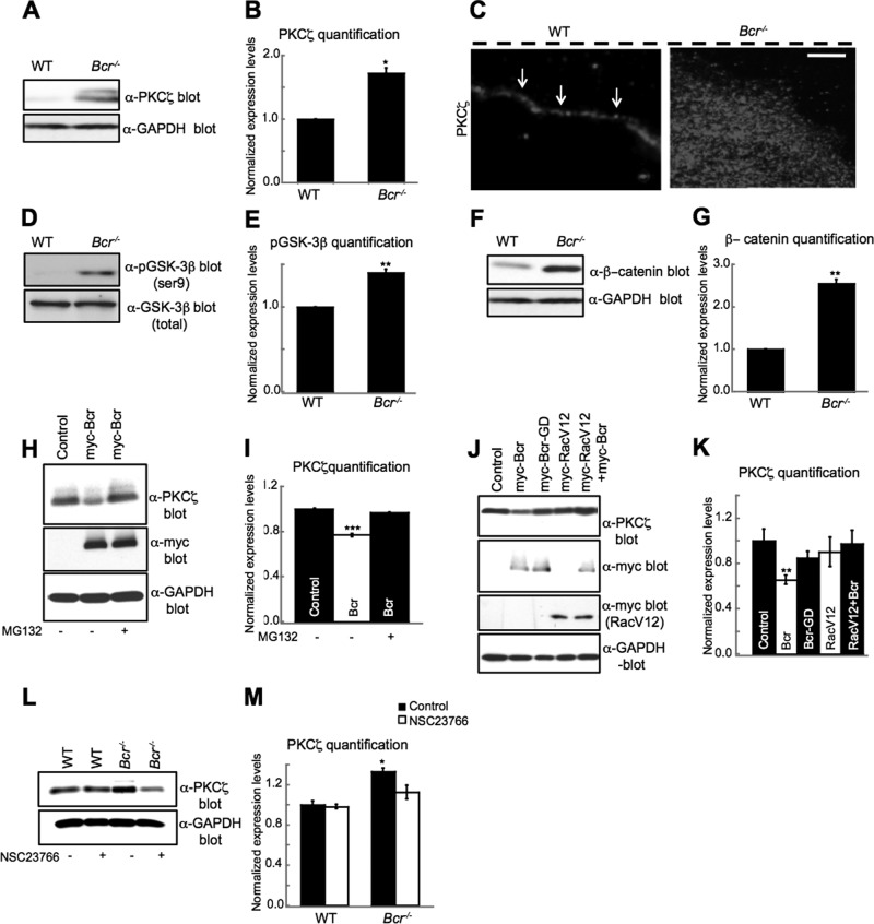
FIGURE 4:
Bcr negatively regulates PKCζ signaling by facilitating its degradation. (A) Western blot analysis of lysates obtained from WT and Bcr−/− mouse cortical astrocytes. Bcr-deficient astrocytes showed an increase in total PKCζ levels. Lysates were also immunoblotted with α-GAPDH antibodies for a loading control. (B) Quantification of PKCζ levels. N = 3. (C) Representative images showing the localization of PKCζ in WT and Bcr−/− astrocytes migrating in a scratch assay. PKCζ is localized to the leading edge (white arrows) in WT cells, whereas PKCζ immunostaining was increased and more diffuse in Bcr−/− cells. Dashed black line shows scratch location. Scale bar: 10 μm. (D) Western blot analysis of lysates obtained from WT and Bcr−/− mouse cortical astrocytes. Bcr-deficient astrocytes showed an increase in p-GSK-3β levels. Lysates were also blotted with total GSK-3β antibodies for a loading control. (E) Quantification of p-GSK-3β levels. N = 3. (F) Western blot analysis of lysates obtained from WT and Bcr−/− mouse cortical astrocytes. Bcr-deficient astrocytes showed an increase in β-catenin levels. Lysates were also blotted with α-GAPDH antibodies for a loading control. (G) Quantification of β-catenin levels. N = 3. (H) Western blot analysis of lysates from COS7 cells expressing myc (control) or myc-Bcr and treated with DMSO (control) or 10 μM of the proteasomal inhibitor MG132. Bcr overexpression reduces PKCζ levels, which is blocked by treating cells with MG132. Lysates were blotted with α-GAPDH antibodies for a loading control. (I) Quantification of PKCζ levels from (H). N = 3. (J) The ability of Bcr to reduce PKCζ levels depends on its Rac-GAP activity. Lysates from COS7 cells expressing myc (control) or myc-tagged Bcr, GAP-dead Bcr (BcrGD), constitutively active Rac (RacV12), or RacV12 plus myc-Bcr were immunoblotted with an α-PKCζ antibody. Lysates were also blotted with α-GAPDH antibodies for a loading control. (K) Quantification of PKCζ levels from (J). N = 3. (L) Bcr reduces PKCζ levels in a Rac-dependent manner. Western blot analysis of lysates obtained from WT and Bcr−/− cortical mouse astrocytes treated overnight with PBS (control) or 50 μM of the Tiam1/Rac inhibitor NSC23766. Lysates were blotted with α-GAPDH for a loading control. (M) Quantification of PKCζ levels from (L). N = 3. Data are shown ± SEM.

