
FIGURE 6:
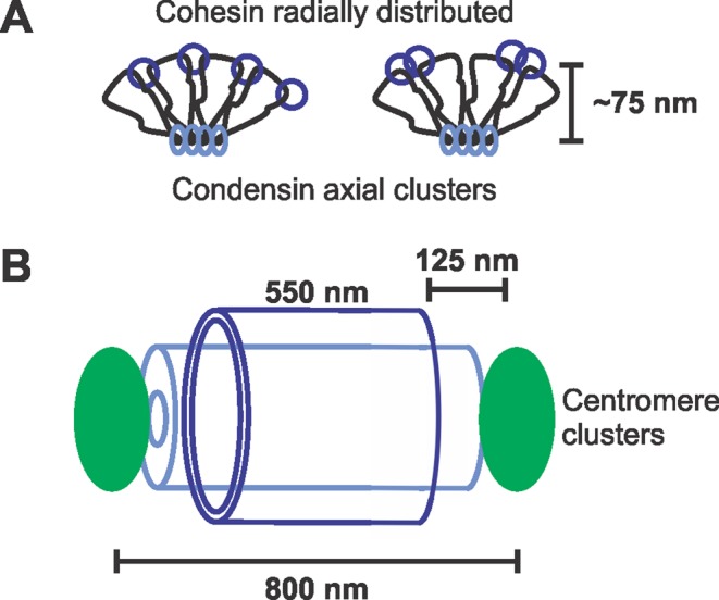
Model of cohesin and condensin in the pericentromere. (A) Condensin (light blue) is localized along the spindle axis in clusters, where it forms rosette-like loops through multiple condensin working cooperatively. Cohesin (dark blue) is localized radially, where it promotes looping to resist outward pulling forces from spindle microtubules. Cohesin is shown as two possible configurations: a single complex (left) or two complexes (right; see review in Haering and Jessberger, 2012). (B) Diagram of the intercentromere region. While condensin can span the length between sister centromere clusters, cohesin is displaced from the centromere cluster by ∼125 nm.