
Figure 3.
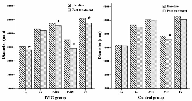
Echocardiography data (mean ± standard deviation) of the intravenous immunoglobulin (IVIG) and control groups at baseline and post-treatment (4 weeks). The diameter of the left atrium (LA), left ventricular end-diastolic diameter (LVDD), left ventricular systolic diameter (LVDS) and diameter of right ventricle (RV) of the IVIG group diminished post-treatment. The diameter of LVDS of the control group diminished. The data of left ventricle (LV) and LA were measured on parasternal long-axis view, and the data of RV and right atrium (RA) were measured on apical four-chamber view. *P<0.05 vs. baseline value.