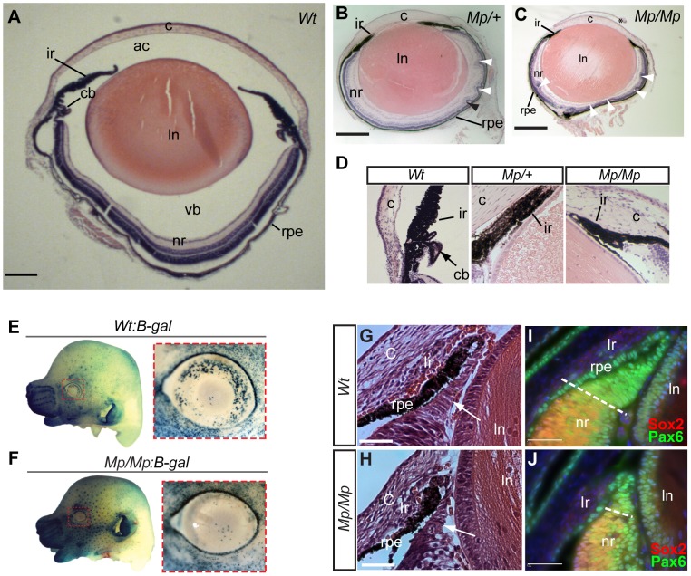
Figure 2. Mp eyes displayed structural defects and abnormal ciliary development.
Histological comparison of eye tissues in Wt (A), Mp/+(B), and Mp/Mp (C) at P21 revealed severe pan-ocular defects. Mutant eyes displayed microphthalmia and retinal rosetting (arrowheads), together with loss of vitreous in the anterior chamber and between lens and retina. Lens size was also reduced in both mutant genotypes. Scale bars = 500 µm; sections are oriented in the sagittal plane. (D) Enlarged view of iris and the anterior region of retinas revealed the absence of ciliary body structures in both mutant types. (E–F) Genetic crosses of Mp with the Wnt-signalling reporter mouse line BAT-gal, revealed a significant reduction in galactosidase-positive cells in E14.5 Mp retinas compared to Wt, specifically in the dorsal and temporal regions of the anterior retina (n = 8 per genotype). In contrast, non-ocular tissue displayed slightly increased staining in Mp:B-gal compared to Wt:B-gal, due to the slight increase in staining time in these samples. Ventral regions had low expression in both genotypes and were used as reference for quantitative comparison (Figure S2). (G–H) Histological analysis of anterior retinas at E15.5 revealed a reduction in non-pigmented ciliary body tissue (arrows) in Mp/Mp compared to Wt. (I–J) Immunohistochemical staining of anterior retinal structures with antibodies specific for Pax6 and Sox2 at E15.5 revealed reduction in the Pax6-positive and Sox2-negative non-pigmented ciliary epithelial region in Mp. Scale bars, 50 µm. The asterisk in C indicates artefactual disruption to the corneal epithelium during sample processing. Abbreviations: ac, anterior chamber; c, cornea; cb, ciliary body; ir, iris; ln, lens; nr, neural retina; rpe, retinal pigmented epithelium; vb, vitreous body.

