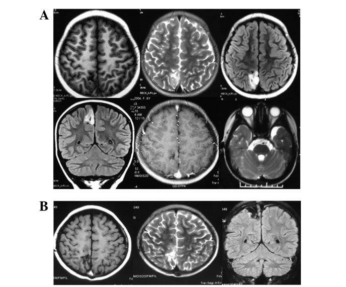
Figure 1.

(A) Preoperative T1-weighted image (WI) showed a low signal, while fluid-attenuated inversion recovery (FLAIR) scanning showed a high signal and enhanced scanning revealed no signals. An arachnoid cyst was visible in the left temporal region. (B) At the six-month postoperative review there was no tumor recurrence.
