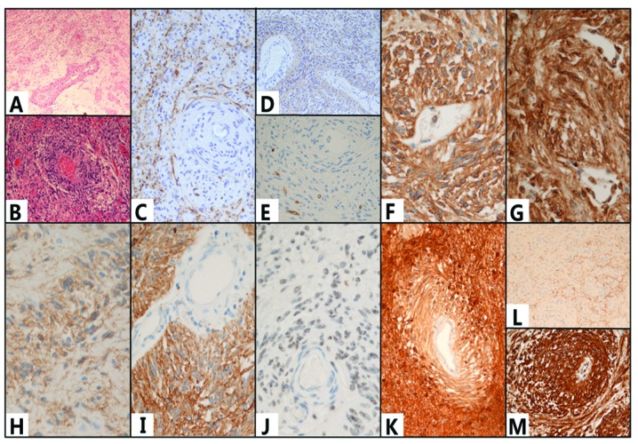
Figure 3.
(A and B) Hematoxylin and eosin (H&E) staining showed that a large number of bipolar cells were clustered around and growing along the long axis of the the blood vessel. The cells were positive for human leukocyte differentiation antigen 34 (CD34) (E), CD99 (F) and D2-40 (G), glial fibrillary acidic protein (GFAP) (I), S-100 protein (K) and vimentin (M); however, they were negative for the neuron markers synaptophysin (Syn) (L) and chromaffin protein/NeuN. There was no expression of microtubule-associated protein 2 (MAP2) (C), nestin (D), epidermal growth factor receptor (EGFR) or (H) oligodendrocyte transcriptor-2 (Olig-2). The was expression of (J) epithelial membrane antigen (EMA) but no neurofilament (NF) in the tumor cells. (A) Magnification, ×10; (B–D,K,L) magnification, ×20; (E–J,M) magnification, ×40.

