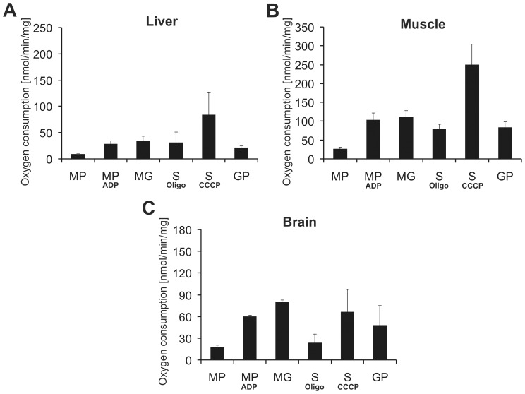
Figure 3. Liver, muscle and brain mitochondria isolated using anti-TOM22 magnetic beads show reliable mitochondrial function.
Oxygen consumption of isolated A liver, B muscle and C brain mitochondria were measured by a Clark electrode. Malate+pyruvate (MP) and malate+glutamate (MG) served as specific substrates for complex I, succinate (S, for complex II and glycerol-3-phosphate (GP) for complex III, respectively. ADP was given to malate+pyruvate to determine state III respiration via complex I. Oligomycin and CCCP were used with succinate to assess state IVo and uncoupled respiration, respectively. The given oxygen consumption units are nmol oxygen/min/mg mitochondria. Data are represented as means of three independent experiments ± SD.

