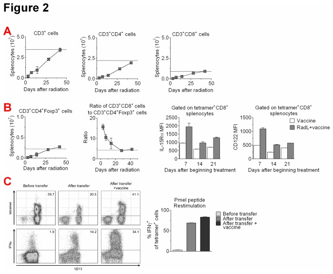
Figure 2. Irradiation followed by lymphocyte infusion leads to marked T cell populations and increased frequency of IFNy+ tumor-antigen specific CD8+ T cells.
A) CD3, CD4 and CD8-expressing splenocytes were quantified by flow cytometry at the indicated time points following irradiation and lymphocyte infusion. Typical numbers from normal mice are indicated by the dotted line. Results shown from one of two experiments with similar results. B) Regulatory T cell splenocytes (CD3+CD4+Foxp3+) and the CD8+ T cell to regulatory T cell ratio were quantified by flow cytometry. Expression of IL-15Rα and CD122 on tetramer+ CD8+ T cells was also quantified by flow cytometry. Results shown from one of two experiments with similar results. C) Mice were irradiated, received an infusion of 30×106 splenocytes from naïve Pmel mice, which express a CD8+ TCR transgene recognizing the melanoma antigen gp100, and immunized weekly with hgp100 DNA plasmid vaccine for 3 doses. Naive Pmel splenocytes, and splenocytes isolated 20 days after infusion into irradiated animals +/- vaccination were restimulated with gp10025-33 peptide (1μg/ml) and evaluated for staining with Pmel-specific tetramer and expression of IFNγ by flow cytometry.

