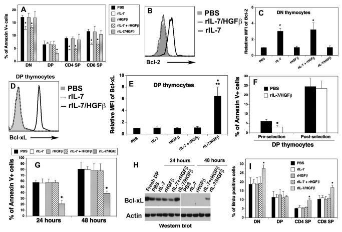
Figure 2. The survival of pre-selection DP and the proliferation of DN and SP thymocytes were enhanced by rIL-7/HGFβ.
(A-F, I) Lethally irradiated BALB/c mice were injected with TCD-BM from B6 mice and treated with cytokines as in Figure 1. On day 30 after BMT, (A) The percentages of Annexin V+ cells in each thymocyte subset were analyzed by flow cytometry. (B) Representative histograms of intracellular staining for Bcl-2 by DN thymocytes. (C) Relative mean fluorescence intensity (MFI) + SD of Bcl-2 by DN thymocytes from the cytokine-treated BMT mice. (D) Representative histograms of intracellular staining for Bcl-xL by DP thymocytes. (E) Relative MFI of Bcl-xL by DP thymocytes. (F) rIL-7/HGFβ treatment enhances the survival of CD69- pre-selection DP thymocytes. (G, H) rIL-7/HGFβ maintains the survival and the expression of Bcl-xL of DP thymocytes in vitro. Purified DP thymocytes were cultured in the presence of equimolar amounts of rIL-7/HGFβ (30 ng/ml), rIL-7 (10 ng/ml) and/or rHGFβ (20 ng/ml), or PBS for 24 and 48 hours. Cells were analyzed for (G) annexin V+ cells by flow cytometry and (H) the expression of Bcl-xL by Western blot. (I) the cytokine-treated allo-BMT mice were injected i.p. with BrdU. The percentages of BrdU+ cells by thymocyte subsets were then analyzed by flow cytometry 4 hour later. The data are representative of 2 independent experiments with 4-6 mice per group. * P<0.05 compared with PBS-treated mice.

