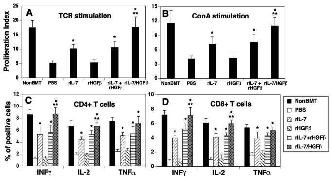
Figure 5. Peripheral T cells in the rIL-7/HGFβ-treated allo-BMT recipients are functional.
Lethally irradiated BALB/c mice were injected with TCD-BM from B6 mice and treated with cytokines as in Figure 1. On day 30 after BMT, (A) splenic CD3+ T cells were stimulated with anti-CD3 and anti-CD28 antibodies (5 μg/ml), and cell proliferation was determined by BrdU incorporation 4 days later. (B) splenic CD3+ T cells were stimulated with Con A (4 μg/ml) and cell proliferation was determined by BrdU incorporation 4 days later. (A and B) Data are shown as stimulation index. (C and D) Splenocytes were stimulated with phorbol myristate acetate and ionomycin, and stained with antibodies for cell surface markers and intercellular cytokines (H-2b, CD4, CD8, IL-2, IFN-γ, and TNFα). The percentage of IL-2, IFN-γ and TNFα positive cells in donor-origin CD4+ and CD8+ T cells was determined by flow cytometry. The data are representative of 2 independent experiments with 4-6 mice per group. * P<0.05 compared with PBS-treated mice. ** P<0.05 compared with rIL-7 and/or rHGFβ-treated mice.

