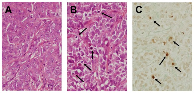
Figure 4. Photomicrograph of subcutaneous human HCC tumor in nude mice that was developed after the injection of HAK-1B cells.

(A) A control mouse that received culture medium alone. The tumor shows a compact arrangement of tumor cells and a sinusoid-like structure in the stroma. (B) A mouse that received a s.c. injection of 0.06 μg of PEG-IFN-α2a. There are some apoptotic tumor-cells characterized by shrinkage and eosinophilic change in the cytoplasm, chromatin condensation and/or fragmentation of nuclei (arrows, HE staining, X200). (C) The same tumor as shown in (B). There are some TUNEL-positive cells showing brown nuclei (arrows, stained by the TUNEL technique, X200).
