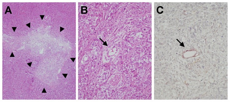
Figure 5. Photomicrograph of resected HAK-1B tumor.
(A) Tumor cells are replaced with large granulation tissue at the middle of resected tumor. (arrowheads, HE staining, X20). (B) Artery-like blood vessels in the tumor (arrow, HE staining, X200). (C) Artery-like blood vessel in the tumor (arrow, CD34/α-SMA double-immunostain, X200).

