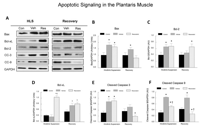
Figure 10. Apoptotic signaling proteins in plantaris muscles.
Plantaris muscle samples were homogenized and total muscle lysates were separated on a 4-12% gradient polyacrylamide gel by routine SDS-PAGE. The proteins were electroblotted to nitrocellulose membranes and the signals were developed by chemiluminescence. A. Representative western blots of apoptotic proteins. Bax, Bcl-xL, Bcl-2, cleaved caspase 3 (CC-3), cleaved caspase 9 (CC-9) and GAPDH. The digital images were quantified as optical density x band area using ImageJ software and normalized to GAPDH, which was used as the loading control for each lane. The data are expressed as the protein signal to the GAPDH signal and are reported as mean ± SEM in arbitrary units. The western blots were run a minimum of three times for each protein, and the data were averaged for each animal’s data point. These data include: Bax (B) Bcl-2 (C), Bcl-xL (D), cleaved caspase 3 (E), and cleaved caspase 9 (F). * P<0.05 vs. cage control; † P<0.05 vehicle vs. resveratrol.

