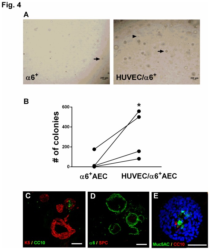
Figure 4. Co-culture of α6β4+ cells with HUVECs ex vivo enhances proliferation but does not alter differentiation.
(A, B) α6+ cells were isolated from normal human distal lung, mixed with HUVECs, and cultured in Matrigel for two weeks. (A) Phase contrast images of α6+ cells cultured without HUVECs (left panel) or with HUVECs (right panel). Arrow indicates colonies arising from α6β4+ cells; arrowhead indicates single HUVEC cells. Scale bar= 200 µm. (B) Colony forming efficiency was analyzed by counting the number of colonies present 14 days after seeding 5000 α6+ cells in the presence or absence of HUVECs. * P<0.05 compared to α6β4+ cells alone. Data are representative of 4 experiments from 4 independent donors. Lines denote paired samples from the same donor. (C) Merged image of colonies arisen from co-culture of α6β4+ cells and HUVEC stained with K-5 (red) and CC10 (green). Scale bar=100 µm. (D) Merged image of immunostaining with SPC (red) and α6 (green) in colonies arisen from co-culture of α6β4+ cells and HUVEC. Scale bar=100 µm. (E) Merged image of immunostaining with goblet cell marker Muc5AC (green) and CC10 (red) in colonies arisen from co-culture of α6β4+ cells and HUVEC. Nuclei were stained with DAPI (blue). Scale bar=50 µm.

