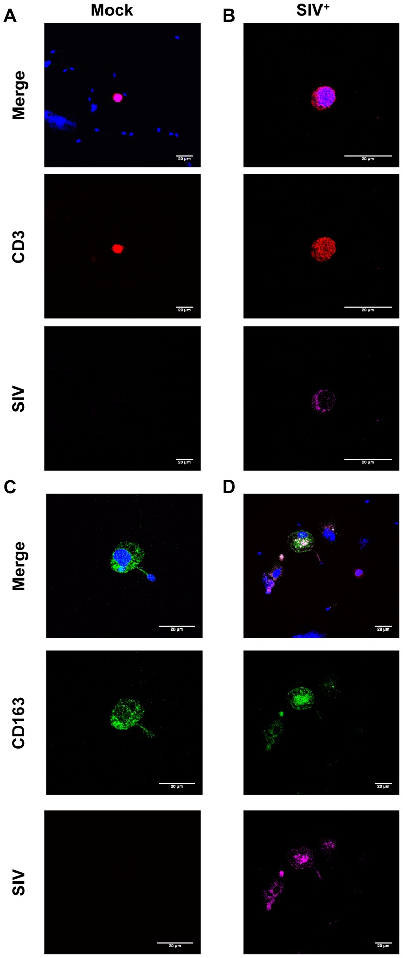
Figure 4. SIV antigens in semen CD4+ T cells and macrophages.
(A–D) Immunocytofluorescence staining targeting CD3 (red), CD163 (green) and SIV Nef (pink) on cytospun CD45+-enriched semen cells from SIV+ (B,D) and uninfected macaques (A,C). Nuclei are stained using DAPI (blue), visible on merged figures (B) SIV+ macaque at 28 dpi. (D) SIV+ macaque at 65 dpi.

