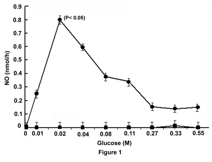
Figure 1. Stimulation of NO synthesis by glucose in mice liver membrane and its inhibition by NAME.
The liver membrane in HEPES buffer, pH 7.4, was prepared from the liver of adult mice as described in the Materials and Methods and incubated with different amounts of glucose in the presence of 2.0mM CaCl2 in a total volume of 1.0ml for 30 min at 37°C. The synthesis of NO was determined by methemoglobin method as described in the Materials and Methods. Identical reaction mixtures contained 0.1mM NAME were incubated under identical conditions and the synthesis of NO was similarly determined.
Each point represents mean ± S.D. of 5 experiments by using 5 different animals each in triplicate.
The solid circles (●) represent the synthesis of NO in the liver membrane preparation in the presence of glucose alone, the solid squares (■) represent the synthesis of NO in the liver membrane preparation in the presence of both glucose and NAME.

