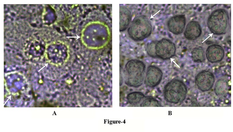
Figure 4. The appearance of Glut-4 on membrane peripheries of hepatocytes incubated with glucose with and without NAME.
The grated liver suspension in HEPES buffer, pH 7.4, was incubated with 0.02M glucose in the presence and absence of 0.1mM NAME in the incubation mixture for 30 min at 37°C. After incubation GLS “chunks” were sliced into 6-8 nm sections by using a cryostat. The sliced sections were treated with Glut-4 antibody to demonstrate the presence of Glut-4 by immunohistochemistry as described in the Materials and Methods.
Panel-A: The immunohistochemistry of liver sections was incubated in the presence of 0.02M glucose. Glut-4 in the liver cells was determined by using fluorescent tagged Glut-4 antibody. White arrow indicates the translocation of Glut-4 transporter to the periphery of liver cell membrane.
Panel-B: The immunohistochemistry of liver sections was incubated in the presence of both 0.02M glucose and 0.1mM NAME by using fluorescent tagged Glut-4 antibody as described in the case of panel-A. White arrow indicates the absence of translocation of Glut-4 transporter to the membrane periphery.
The figures shown are typical representative of six different experiments using 6 different animals.

