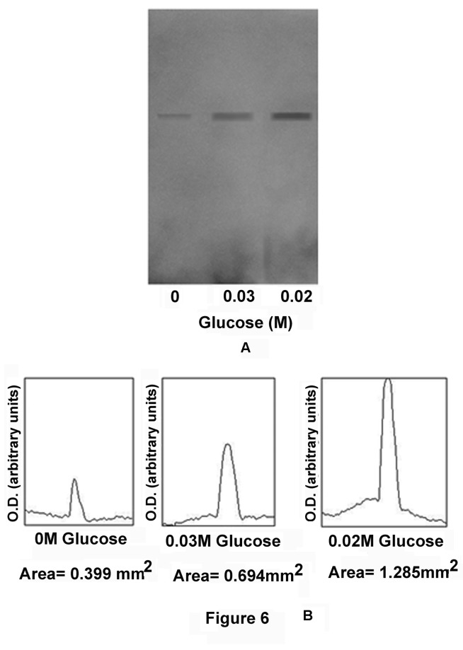
Figure 6. Immunoblot analysis of Glut-4 synthesis by in vitro translation of Glut-4 mRNA in the GLS.
The grated liver suspension in HEPES buffer, pH 7.4, was incubated with different concentrations of glucose for 30 min at 37°C. After incubation, nucleic acids which contained mRNA of Glut-4 were extracted and translated in vitro as described in the Materials and Methods. The reaction supernatants were subjected to immunoblot analysis using Glut-4 antibody (Panel-A), the immunopositive bands were quantitated by using Image-J program by computer analysis. The integrated area of each band was also calculated (Panel-B).
Panel-A: Immunopositive bands of Glut-4 synthesis in GLS incubated in the presence of different concentrations of glucose in the incubation mixture as indicated.
Panel-B: Integrated area of each of the immunopositive band as shown in the panel-A.
Both Panel-A and Panel-B are the representative of experiments by using 6 different animals.

