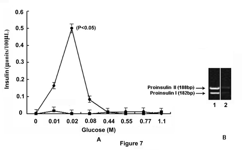
Figure 7. Role of NO in insulin synthesis in glucose treated GLS via the expression of proinsulin genes.
The grated liver suspension in HEPES buffer, pH 7.4, was incubated with different amounts of glucose as indicated for 30 min at 37°C. After incubation, nucleic acids were extracted and insulin mRNA was translated in vitro as described in the Materials and Methods. The amount of insulin synthesized, was quantitated by ELISA using anti-insulin antibody. In parallel experiments, the incubation mixture was incubated with glucose and 0.1mM NAME and the synthesis of insulin was similarly determined.
Panel-A: Glucose induced synthesis of insulin in the presence and absence of NAME. The solid circles (●) represent insulin synthesis in the presence of different amounts of glucose and the solid squares (■) represent the synthesis of insulin in the presence of both glucose and 0.1mM NAME.
Panel-B: Agarose gel electrophoresis of cDNA prepared from the isolated insulin mRNA.
‘1’ and ‘2’ represents the expression of proinsulin genes I and II in the presence and absence of 0.02M glucose respectively.
Results shown are representatives of the optical densities obtained from 6 different experiments using 6 different animals.

