
Figure 3.
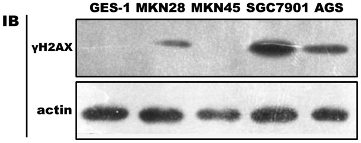
Expression of γH2AX in non-cancerous and cancerous gastric cell lines measured by western blotting. Cell lines were grown for two days and then subjected to total cellular protein isolation. γH2AX, phosphorylated H2AX at Ser 139.
Official websites use .gov
A
.gov website belongs to an official
government organization in the United States.
Secure .gov websites use HTTPS
A lock (
) or https:// means you've safely
connected to the .gov website. Share sensitive
information only on official, secure websites.
Expression of γH2AX in non-cancerous and cancerous gastric cell lines measured by western blotting. Cell lines were grown for two days and then subjected to total cellular protein isolation. γH2AX, phosphorylated H2AX at Ser 139.