
Figure 2.
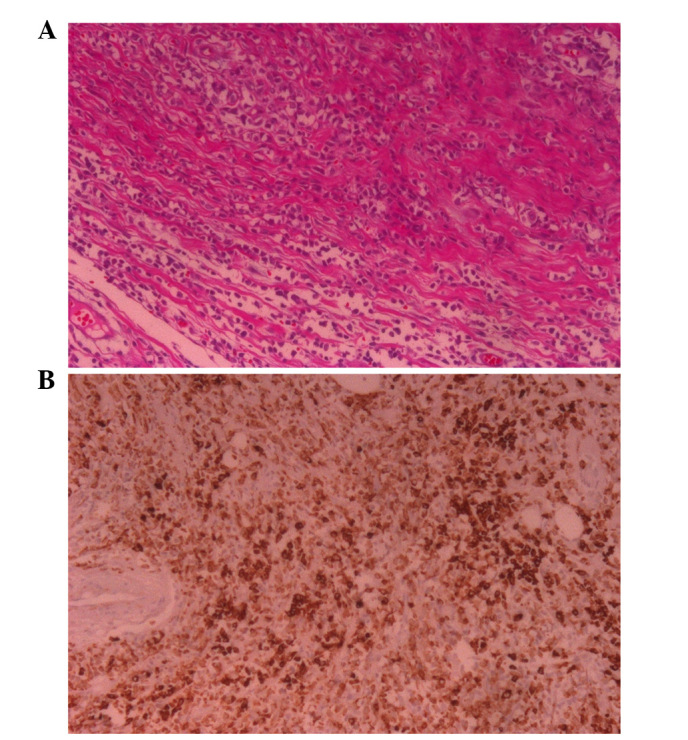
(A) In case 2, mesenchymal tissues were packed into the tumor cells, arranged into a line and the fatty tissue was infiltrated at the edge of the tumor. (B) Immunohistochemistry stain showing that MPO was markedly positive in the cell plasmid. (Hematoxylin and eosin staining; magnification, ×200).