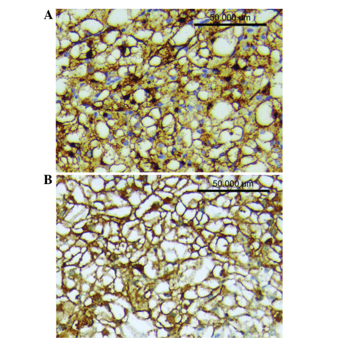
Figure 2.

Immunohistochemical results of hepatic PEComa (x200). (A) Tumor cells showing strong and diffuse positive staining for HMB-45 (+++). (B) Tumor cells showing strong and diffuse positive staining for SMA (+++). PEComa, perivascular epithelioid cell tumor; HMB-45, human melanoma black-45; SMA, smooth muscle actin.
