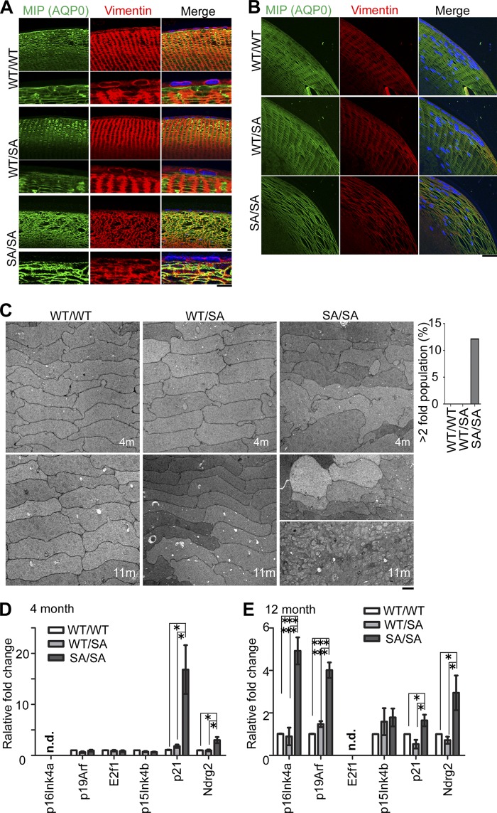
FIGURE 6.
Progeria-like phenotypes apparent in VIMSA/SA lenses. A and B, disorganization of lens fiber cells in VIMSA/SA mice. Anterior (A) or equatorial (B) lens fiber cells from 4-month-old mice were immunostained with anti-MIP (AQP0; green), anti-vimentin (red), and DAPI (blue; nuclei). C, electron micrographs of lens at 4 (upper) or 11 (lower) months of age. The dispersion in the size distribution was evaluated as the percentage of cells >2 larger than the average size of cells (right graph). D and E, mRNA expression of the indicated genes were analyzed through quantitative RT-PCR using the lens of 4-month-old (D) or 12-month-old (E) mice (n = 5 mice per genotype). n.d. means “no detected signals.” Scale bars, 30 μm (A, upper) or 10 μm (A (lower) and B and C). *, p < 0.05; ***, p < 0.001.

