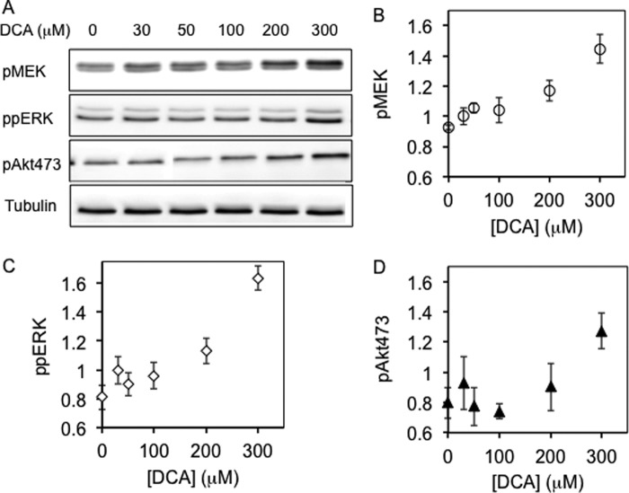
FIGURE 6.
Bile acid enhances MAPK signaling induced by EGF. BHK cells were serum-starved for 2 h, treated with DCA for 1 h, stimulated with 2.5 ng/ml EGF for 5 min, and harvested. Whole cell lysates were used to blot for phosphorylated MEK, ERK, or Akt. A, representative Western blots with antibodies against pMEK, pERK, or pAkt473. B–D, normalized band intensity values (see “Experimental Procedures”) for pMEK (B), ppERK (C), and pAkt (D) in the form of means ± S.E. (error bars) as a function of bile acid concentrations from three independent experiments.

