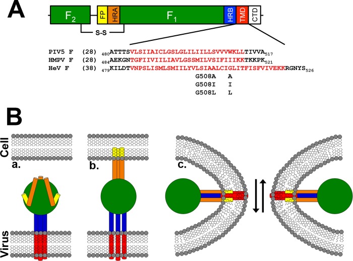
FIGURE 1.
Schematic of a paramyxovirus F protein and model of membrane fusion. A, diagram of a mature paramyxovirus F protein, with TM sequences indicated. FP = fusion peptide; HR = heptad repeat; TMD = transmembrane domain; CTD = cytoplasmic tail. B, model for F protein-promoted membrane fusion. Part a, the HRB domains adjacent to the TM form a coiled-coil in the prefusion state, whereas the HRA domains are present as a series of small helices. Part b, triggering of fusion leads to insertion of the fusion peptide into the target membrane and formation of an HRA coiled-coil, and separation of the HRB helices and TM domains. Part c, fusion pore opening occurs concomitant with formation of a six-helix bundle with the HRA segments on the interior of the bundle, placing the HRB and TM segments on the exterior.

